40 fluorescent labels and light microscopy
Fluorescent Labeling - What You Should Know - PromoCell Fluorescence microscopy allows the identification of cells and cellular components and the monitoring of cell physiology with high specificity. Fluorescence microscopy separates emitted light from excitation light using optical filters. The use of two indicators also allows the simultaneous observation of different biomolecules at the same time. Fluorescence Microscopy - New York Microscope Company Fluorescence microscopy uses a high-intensity light source that excites a fluorescent molecule called a fluorophore in the sample observed. The samples are labeled with fluorophore where they absorb the high-intensity light from the source and emit a lower energy light of longer wavelength.
Fluorescence Microscopy Sir George Stokes, a British scientist, first discovered fluorescence in 1852 when he observed that the mineral fluorite (Fig. 1, molecular formula CaF2) emitted red light when it was illuminated by ultraviolet excitation. Early investigations in the 19th century showed that many specimens (including minerals, crystals, drugs, butter, chlorophyll, and vitamins) fluoresce when irradiated with…

Fluorescent labels and light microscopy
Imaging Flies by Fluorescence Microscopy: Principles ... The development of fluorescent labels and powerful imaging technologies in the last two decades has revolutionized the field of fluorescence microscopy, which is now widely used in diverse scientific fields from biology to biomedical and materials science. ... has brought about the era of fluorescence light microscopy. The first fluorescence ... Fluorescent Labelling - an overview | ScienceDirect Topics Fluorescence microscopy Fluorescent labeling methods are generally based on reactive derivatives of fluorophores that selectively bind to functional groups contained in target biomolecules and are widely used in biotechnology because of their non-destructive properties and the high sensitivity of fluorescence techniques ( Sahoo, 2012 ). A quick guide to light microscopy in cell biology - PMC Fluorescence microscopy uses fluorescent dyes (fluorophores), which are molecules that absorb one wavelength of light (the excitation wavelength) and emit a second, longer wavelength of light (the emission wavelength).
Fluorescent labels and light microscopy. Light Microscope- Definition, Principle, Types, Parts ... A minimum distance (d) between two objects that distinguishes them to be two separate entities, determined by the wavelengths of the light can be calculated by an Abbe equation using the wavelength of the light that illuminated the specimen (Lambda, λ) and the numerical aperture (NA, n sin Ɵ) i.e. d=0.5 λ/n sin Ɵ In Silico Labeling: Predicting Fluorescent Labels in ... Fluorescence microscopy images can be predicted from transmitted-light z stacks • 7 fluorescent labels were validated across three labs, modalities, and cell types • New labels can be predicted using minimal additional training data Summary Microscopy is a central method in life sciences. Fluorescence Imaging - Teledyne Photometrics Fluorescent molecules (known as fluorophores) are used to label samples, and fluorophores are available that emit light in virtually any color. In a fluorescent microscope, a sample is labeled with a fluorophore, and then a bright light ( excitation light) is used to illuminate the sample, which gives off fluorescence ( emission light ). Fluorescence Microscopy vs. Light Microscopy This means that fluorescent microscopy uses reflected rather than transmitted light. For example, a commonly used label is green fluorescent protein (GFP), which is excited with blue light and...
New fluorescent label provides a clearer picture of how ... A molecule of interest is labelled with a special fluorescent dye that flashes on and off like a blinking star. Unlike traditional fluorescence microscopy, which uses labels that glow constantly,... Fluorescent labeling of abundant reactive entities (FLARE ... Fluorescence microscopy is a technique that is commonly used in the biomedical sciences. It offers the powerful ability to visualize structures or molecules in three dimensions within biological... Label-free imaging of live cells - CytoSMART Study live cells using non-invasive & non-toxic methods Live-cell imaging has become a fundamental tool to better understand cellular processes and biological functions. A non-invasive and non-toxic alternative to fluorescent microscopy is label-free imaging. Although the resolution achievable… Fluorescence Microscopy & Cell Imaging | Research | UNM ... The Fluorescence Microscopy and Cell Imaging Shared Resource provides UNM researchers access to state-of-the art instrumentation for multiple fluorescence and transmitted light microscopy techniques: Laser scanning single and multi-photon microscopes and hyperspectral imaging systems enable simultaneous visualization and quantification of ...
Light Microscopy in Trypanosomes: Use of Fluorescent ... Spray a glass microscope slide with 70% (v/v) ethanol and wipe clean. Pipet 3 μL of cell solution onto the glass slide and carefully place a No. 0 coverslip (50 × 22 mm) on top of the cell solution. 6. Check the slide on an inverted microscope using a 20× objective to ensure that the cells are well immobilized on the slide ( see Note 7 ). 7. Fluorescent tag - Wikipedia Fluorescent tag. S. cerevisiae septins revealed with fluorescent microscopy utilizing fluorescent labeling. In molecular biology and biotechnology, a fluorescent tag, also known as a fluorescent label or fluorescent probe, is a molecule that is attached chemically to aid in the detection of a biomolecule such as a protein, antibody, or amino acid. Evanescent Light-Scattering Microscopy for Label-Free ... Measurements and theoretical analysis revealed that light-scattering signals originating from single surface-bound lipid vesicles enable characterization of their sizes without employing fluorescent lipids as labels. Researchers Develop New Fluorescent DNA Label for Clinical ... Superresolution fluorescence microscopy creates clearer biological images through the blinking on and off of specific labels at different times, allowing a composite image to be reconstructed showing all of the labeled components in high resolution.

Buchmann Institute for Molecular Life Sciences - BMLS
Light Sheet Fluorescence Microscopy - an overview ... Applications of single-molecule fluorescence microscopy. (A) The photophysical properties of a fluorophore contain information about its position and its state. This allows, for example, tracking molecules, observing conformational and constitutional changes, or following chemical reactions. (B) Examples for applications in biology and chemistry.

FISH - Semrock
Visualizing the invisible: New fluorescent DNA label ... A molecule of interest is labelled with a special fluorescent dye that flashes on and off like a blinking star. Unlike traditional fluorescence microscopy, which uses labels that glow constantly,...

35 Microscope Drawing With Label - Labels Information List
Label-free prediction of three-dimensional fluorescence ... Fluorescence microscopy can resolve subcellular structure in living cells, but is expensive, slow, and toxic. Here, we present a label-free method for predicting 3D fluorescence directly from transmitted light images and demonstrate its use to generate multi-structure, integrated images.
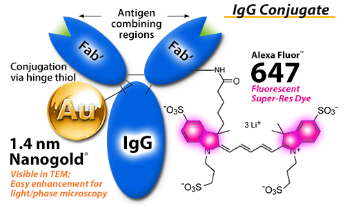
FluoroNanogold™ combined fluorescent and gold nanoparticle immunoprobe
Label-free prediction of three-dimensional fluorescence ... Label-free prediction of three-dimensional fluorescence images from transmitted-light microscopy Understanding cells as integrated systems is central to modern biology. Although fluorescence microscopy can resolve subcellular structure in living cells, it is expensive, is slow, and can damage cells.

Development a flexible light‐sheet fluorescence microscope for high‐speed 3D imaging of calcium ...
Label-free prediction of three-dimensional fluorescence ... We present a label-free method for predicting three-dimensional fluorescence directly from transmitted-light images and demonstrate that it can be used to generate multi-structure, integrated...

(a) Tapping-mode AFM image of purified 10 nm NP-A-633 after three... | Download Scientific Diagram
Fluorescence Microscope: Principle, Types, Applications ... Fluorescence microscopy is a type of light microscope that works on the principle of fluorescence. A substance is said to be fluorescent when it absorbs the energy of invisible shorter wavelength radiation (such as UV light) and emits longer wavelength radiation of visible light (such as green or red light).
Post a Comment for "40 fluorescent labels and light microscopy"